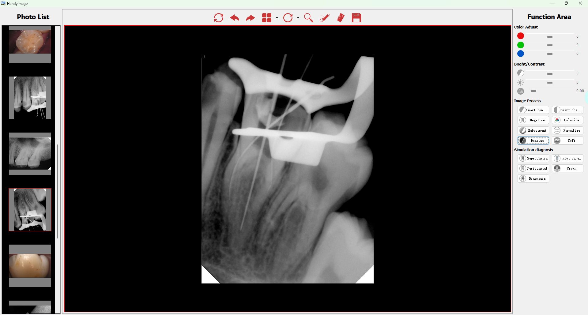
The application of photography in dental pathology dates back to 1840, when the first dental school opened alongside the world’s first photographic gallery.1 Dentists R. Thompson and W. Elde were the first to document before-and-after intraoral images of dental procedures in 1848, paving the way for future advancements in dental diagnosis and therapy, such as the inception of the HD intraoral camera (IOC).
I have worked hand in hand in these two fields, with clinical photography becoming an essential aspect of a patient’s medical record and treatment strategy. Dental decisions are now nearly impossible to make without using an IOC. IOCs have grown in importance as valuable tools for presenting information, record keeping, and patient education. Sensor Dental

In the last couple of decades, most dental patients have seen images of the inside of their mouths as cameras of various types have become a necessity in practically every dental office, regardless of specialty. However, many years ago this was not the case, when only x-rays and scans with images that were incomprehensible to patients were the norm. More than half a century ago, dentists began searching for a camera that would allow patients to see precisely what dentists see.
In the 1970s, instant film cameras became the solution for obtaining immediate photographic results. Then, in the 1980s, computers began to play a more prominent role in dentistry. New, groundbreaking IOCs coupled with video features allowed patients to look inside their mouths in real time, together with the dentist.2
Nevertheless, early dental IOCs were cumbersome. They took up a lot of room and were expensive, costing up to $40,000 per unit. These days, complete IOC systems cost just a fraction of the price, with some being only a few hundred dollars. Bulky docking stations have been replaced with USB connections that are lighter, easy to use, and produce better quality photographs. As a result of these significant developments in dental technology, the IOC is now a routine part of every dental practice.
For years, dental professionals have had difficulty describing a patient’s clinical situation as well as justifying proposed treatment plans. There was always some degree of insecurity when there was no physical evidence to back up the dentist’s claims. IOCs offer visual transparency, allowing dentists and patients to communicate more effectively. Patients can now see what their dentists see—accurate, enlarged details of their teeth and surrounding structures without any pain or invasiveness. Patients are involved in the process, enabling them to completely comprehend what is going on and why the recommended treatment is necessary.
Instant image deletion and collection makes intraoral imaging a valuable tool during consultations. Dental professionals and patients can visually evaluate previous images, take new ones, and compare changes, subsequently minimizing patient anxiety and facilitating a trustworthy patient-doctor relationship.
High-definition (HD) images are viewed on television every day, but intraoral cameras have only recently been able to take advantage of this substantial improvement in image quality. The IOC’s ability to produce HD images took several years to perfect, and today we are finally able to achieve real HD picture quality.
But what exactly does HD entail? Although some cameras boast “high-definition,” they only have a 720p resolution. The “p” in 720p or 1080p stands for progressive scanning, which updates full-frame images more quickly than traditional interlaced images. A true HD camera, such as the MouthWatch Plus+, will deliver a 1080p image that will capture 1,152,000 more pixels than a 720p image, resulting in an increase of more than 50% in resolution.3 Higher resolution means more pixels per inch (PPI), which means more pixel information and thus a higher-quality, sharper image.
Consider pixels in the same way that you would a set of building blocks. The more pixels you have, the more you have to work with when building an image. This increased number of pixels results in a sharper, more realistic video and still images. The additional pixels improve both video and still image quality.
Another crucial factor is the rate at which intraoral images are transmitted to a computer monitor, especially when the camera is moving within the mouth. Anything lower than 30 frames per second will cause the images to appear “jerky”; 60 frames per second is necessary for the smoothest video display.
Patients’ dental anatomy can be documented in extraordinary detail by utilizing high-definition cameras that produce sharp, high-resolution images. Today, HD IOCs can produce super-macro close-ups of up to 80 times, including photographs of individual teeth as well as whole arch and patient portraits. These options allow us to make better diagnoses in daily clinical operations, from showing patients dramatically magnified periodontal concerns to examining leaky fillings or reviewing margins on a newly constructed lab crown.
Anatomically accurate color and high-definition resolution are essential in dental imaging because they assist in capturing fine features that can be used to monitor the oral cavity, explain specific problems to patients, and even support insurance claims. Color can reveal hidden abnormalities and indicate regions that would otherwise be easily overlooked in the early stages, including examining for oral lesions, oral cancer concerns, or gingival irritation.
Dental digital photography has evolved into a convenient and straightforward method for documenting patients’ dental pathology and anatomy. Intraoral images allow patients to witness the condition of their teeth and gums from the dentist’s point of view, which aids understanding of the rationale behind a proposed treatment. Digital images are also convenient to retain for future legal or academic purposes. As a result, a digital IOC or an HD IOC should be essential equipment in every dental practice.

Camera Intraoral Com Monitor Editor’s note: This article first appeared in Through the Loupes newsletter, a publication of the Endeavor Business Media Dental Group. Read more articles and subscribe to Through the Loupes.